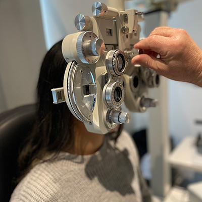
The Benefits of Retinal Imaging
What is an Optomap?
Optomap is an ultra-widefield retinal examination.
With traditional, small-field, and even widefield retinal imaging, only 10-100⁰ of the retina can be captured in a single image. Optomap is the only true, clinically validated, ultra-widefield retinal image than can capture 82% or 200⁰ of the retina, in a single capture, in each imaging modality. An increase of 50% over the next closest imaging device. With Optomap auto-montage, up to 97% or 220⁰ of the retina can be imaged with the multi-capture, montaging functionality.

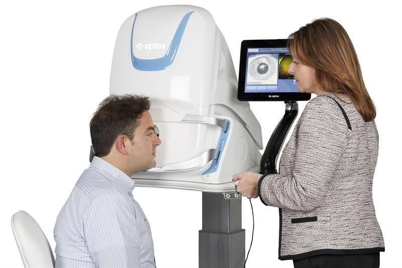 Optomap offers unparalleled views of the retina which provide eye care professionals like us, with the following:
- The only anatomically correct 200⁰ or 82% image of the retina
- Multiple retinal imaging modalities to see more, discover more, and treat more patient diseases and pathologies, more effectively
- More than 1000 published and ongoing clinical trials, as well as thousands of case studies and testimonials. All showing the long-term value of Optomap imaging in diagnosis, treatment planning and patient engagement
 What Retinal Complications Can Optomap Detect?
An Optomap image provides a bigger picture and more clinical information which facilitate the early detection, management and effective treatment of disorders and diseases evidenced in the retina. Including retinal detachments and tears, glaucoma, diabetic retinopathy and age related macular degeneration. Retinal imaging can also indicate evidence of systemic diseases such as hypertension and certain cancers.
Please see ‘A Retinal Reference Guide’ image for different types of issues that can occur with the retina that Optomap allows us to effectively diagnose.
Optomap VS Dilation
Without the vast benefits of the Optomap, the next best solution to increase visibility of the retina and any potential complications occurring inside the eye, is to complete a dilated retinal exam.
This involves using an eye drop to relax the iris (the coloured part of the eye), increasing the size of the pupil which then becomes unresponsive to light (does not contract). This allows the doctor to see a great deal more inside of the eye using either the slit lamp or OCT (optical coherence tomography) imaging machine.
Dilating a patients pupils means that they cannot drive for at least 3-5 hours afterwards. The drops make vision light sensitive and blurry. Please see the list of benefits for using Optomap instead of dilation:
- Much faster appointment with Optomap. 5 minutes compared to 45 minutes (waiting for the drops to take action can take some time)
- The patient can drive to the appointment and vision will not be blurry or light sensitive
- Easy option to compare images over time through the advanced Optos software. This is especially advantageous for our patients undergoing intravitreal injections for one of the many names retinal diseases above
- Green / red light lasers that are used during the Optomap scan allows the doctor to see and enhance a variety of retinal disorders making treatment planning much more effective and accurate
|By: Paula
**Please note this blog is not a substitute for medical advice. If you have any concern about your vision quality or eye health, we urge you to contact your GP or Ophthalmologist**